
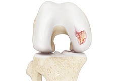
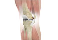
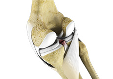
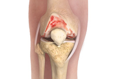
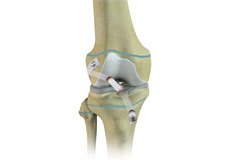
Knee Arthritis
The joint surface is covered by a smooth articular surface that allows pain-free movement in the joint. Arthritis is a general term covering numerous conditions where the joint surface or cartilage wears out. This surface can wear out for several reasons; often the definite cause is not known.
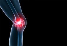
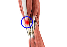
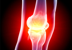
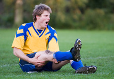
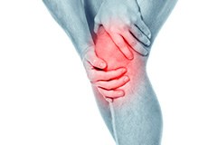
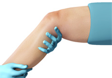
Knee Pain
Knee pain is a common condition affecting individuals of various age groups. It not only affects movement but also impacts your quality of life. An injury or disease of the knee joint or any structure surrounding the knee can result in knee pain.
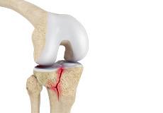
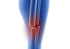
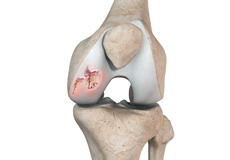
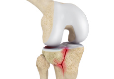
Knee Fracture
A fracture is a condition in which there is a break in the continuity of the bone. In younger individuals, these fractures are caused by high energy injuries, as from a motor vehicle accident. In older people, the most common cause is a weak and fragile bone.
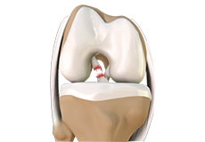
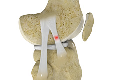
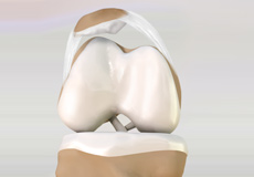
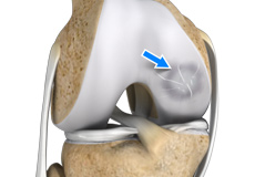
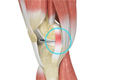
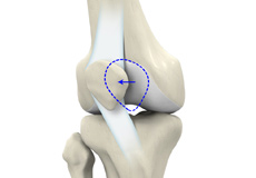
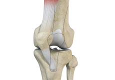
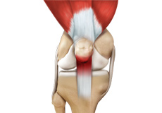
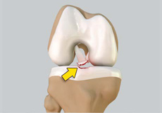
ACL Tears
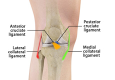
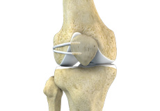
The anterior cruciate ligament (ACL) is one of the major ligaments of the knee. It is located in the middle of the knee and runs from the femur (thighbone) to the tibia (shinbone). The ACL prevents the tibia from sliding out in front of the femur. Together with the posterior cruciate ligament (PCL), it provides rotational stability to the knee.
Multiligament Knee Injuries
Injury to more than one knee ligament is called a multiligament knee injury and may occur during sports or other physical activities.
Knee Sprain
Knee sprain is a common injury that occurs from overstretching of the ligaments that support the knee joint. A knee sprain occurs when the knee ligaments are twisted or turned beyond its normal range, causing the ligaments to tear.
Patellofemoral Instability
Patellofemoral instability means that the patella (kneecap) moves out of its normal pattern of alignment. This malalignment can damage the underlying soft structures such as muscles and ligaments that hold the knee in place.
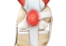
Kneecap Bursitis
Bursitis refers to the inflammation and swelling of the bursa. Inflammation of the bursa in front of the kneecap (patella) is known as kneecap bursitis or prepatellar bursitis.
Chondromalacia Patella
Chondromalacia patella is a common condition characterized by softening, weakening and damage of the cartilage. The condition is most often seen in young athletes and older adults who have arthritis of the knee. It especially occurs in women.
Iliotibial Band Syndrome
Iliotibial band syndrome is an overuse injury resulting from the inflammation of the iliotibial band. It occurs when the iliotibial band and the lower outside portion of the thighbone at the knee joint rub against each other.
Lateral Patellar Compression Syndrome
Lateral patellar compression syndrome refers to pain under and around your kneecap. It is a common complaint among runners, jumpers and other athletes such as skiers, cyclists, and soccer players.
Fractures of the Tibia
The lower leg is made up of two long bones called the tibia and fibula that extend between the knee and ankle. The tibia or shinbone is the larger of the two bones. It bears most of the body’s weight and helps form the ankle joint and knee joint.
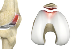
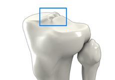
Osteochondritis Dissecans of the Knee
Osteochondritis dissecans is a joint condition in which a piece of cartilage, along with a thin layer of the bone separates from the end of the bone because of inadequate blood supply. The separated fragments are sometimes called “joint mice”.
Knee Injury
Pain, swelling, and stiffness are the common symptoms of any damage or injury to the knee. If care is not taken during the initial phases of injury, it may lead to joint damage, which may end up destroying your knee.
Unstable Knee
Damage to any of these supportive structures causes instability of the knee joint. An unstable knee can be caused by the sudden twisting of the knee, tears of the meniscus, ligament or capsule, osteoarthritis of the knee (wear and tear of the cushioning cartilage tissue between the bones) and sports injuries.
Knee Infection
Knee infection is a serious medical condition that needs immediate treatment. Infection may occur followed by a knee replacement surgery or trauma and is usually caused by bacteria. Infection may spread to the space of the knee joint or deep layers of your knee causing serious complications.
MCL Tears
The medial collateral ligament (MCL) is the ligament located on the inner part of the knee joint. It runs from the femur (thighbone) to the top of the tibia (shinbone) and helps in stabilizing the knee.
MCL Sprains
MCL sprains occur due to a sudden impact from the outside of your knee, most commonly while playing sports such as rugby and football. Rarely, the MCL can get injured when the knee gets twisted or following a quick change in direction.
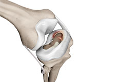
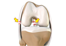
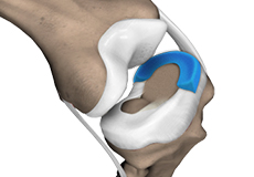
Meniscal Injuries
Meniscal tears are one of the most common injuries to the knee joint. It can occur at any age but are more common in athletes involved in contact sports. The meniscus has no direct blood supply and for that reason, when there is an injury to the meniscus, healing is difficult.
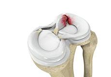
Meniscal Tears
A meniscal tear is a common knee injury in athletes, especially those involved in contact sports. A sudden bend or twist in your knee causes the meniscus to tear. Elderly people are more prone to degenerative meniscal tears as the cartilage wears out and weakens with age.
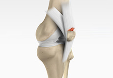
Fractures of the Patella
The patella or kneecap is a small bone present in the front of your knee where the thigh bone meets the shinbone. It provides protection to your knee and attachment to muscles in the front of the thigh. An injury to the knee can result in a break or fracture of the patella.
Ligament Injuries
The most common types of ligament injuries include:
ACL Tear
MCL Tear
PCL Tear
Multiligament Instability
The knee is a complex joint of the body that is vital for movement. The four major ligaments of the knee are anterior cruciate ligament (ACL), posterior cruciate ligament (PCL), medial collateral ligament (MCL) and lateral collateral ligament (LCL).
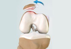
Patellar Dislocation/Patellofemoral Dislocation
Patellar dislocation occurs when the patella moves out of the patellofemoral groove, (trochlea) onto the bony head of the femur. If the kneecap partially comes out of the groove, it is called subluxation; if the kneecap completely comes out, it is called dislocation (luxation).
PCL Injuries
Posterior cruciate ligament (PCL), one of the four major ligaments of the knee, is situated at the back of the knee. It connects the thighbone (femur) to the shinbone (tibia). The PCL limits the backward motion of the shinbone.
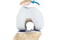
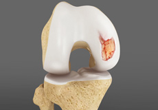
Chondral or Articular Cartilage Defects
The articular or hyaline cartilage is the tissue lining the surface of the two bones in the knee joint. Cartilage helps the bones move smoothly against each other and can withstand the weight of your body during activities such as running and jumping.
Patellar Instability
Any damage to the supporting ligaments may cause the patella to slip out of the groove either partially (subluxation) or completely (dislocation). This misalignment can damage the underlying soft structures such as muscles and ligaments that hold the kneecap in place.
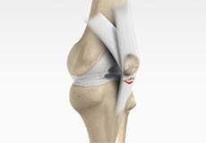
Patella Fracture
The kneecap or patella forms a part of the knee joint. It is present at the front of the knee, protecting the knee and providing attachment to various muscle groups of the thigh and leg. The undersurface of the kneecap and the lower end of the femur are coated with articular cartilage, which helps in smooth movement of the knee joint.
Recurrent Patella Dislocation
The patella (kneecap) is a small bone that shields your knee joint. It is present in front of your knee, on a groove called the trochlear groove that sits at the junction of the femur (thighbone) and tibia (shinbone). Articular cartilage presents below the patella and end of the femur cushion and helps the bones glide smoothly over each other when the legs move.
Quadriceps Tendon Rupture
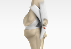
The quadriceps can rupture after a fall, direct blow to the leg and when you land on your leg awkwardly from a jump. Quadriceps tendon rupture most commonly occurs in middle-aged people who participate in sports that involve jumping and running.
Patellar Tendon Rupture
The patellar tendon works together with the quadriceps muscle and the quadriceps tendon to allow your knee to straighten out. Patella tendon rupture is the rupture of the tendon that connects the patella (kneecap) to the top portion of the tibia (shinbone).
Lateral Meniscus Syndrome
Lateral meniscus syndrome is characterized by an injury caused by the tearing of the cartilage tissue or a rare case of a congenital abnormality called a discoid meniscus, which results in knee pain.
Tibial Eminence Spine Avulsion Fracture
Tibial eminence spine avulsion fracture is the avulsion (tearing away) of the tibial eminence.
Posterolateral Instability
Posterolateral instability, also known as posterolateral rotatory instability (PLRI), is a common pattern of knee instability that results from injuries to the structures that support the outside of the knee joint, the posterolateral corner. When individuals injure the knee ligaments, they are most likely to injure the structures of the posterolateral corner.
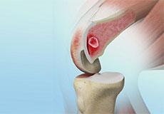
Osteochondral Defect of the Knee
An osteochondral defect, also commonly known as osteochondritis dissecans, of the knee refers to a damage or injury to the smooth articular cartilage surrounding the knee joint and the bone underneath the cartilage. The degree of damage may range from a rupture of the cartilage to a slight crack of the bone to a piece of the bone breaking off within the joint.
Articular Cartilage Injury
Tibial eminence spine avulsion fracture is the avulsion (tearing away) of the tibial eminence.
Tibial Shaft Fracture
A tibial shaft fracture is a crack or break in the middle section of the tibia bone due to severe trauma.
The lower leg is made up of two long bones called the tibia and fibula that extend between the knee and ankle and help form the ankle joint and knee joint.
Patellar Tracking Disorder/Patellar Maltracking
Patellar tracking disorder, also known as patellar maltracking, is a condition in which the kneecap (patella) moves sideways from its groove when the leg is bent or straightened.
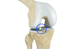
Loose Bodies in the Knee
Loose bodies are fragments of detached cartilage or bone inside the knee joint. These fragments may be free floating (unstable) or may be trapped (stable) within the joint. Depending on the severity, you may have one or more loose bodies in your knee joint.
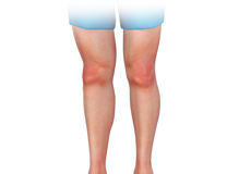
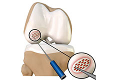
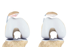
Knee Osteoarthritis
Osteoarthritis also called degenerative joint disease, is the most common form of arthritis. It occurs most often in older people. This disease affects the tissue covering the ends of bones in a joint (cartilage).In a person with osteoarthritis, the cartilage becomes damaged and worn out causing pain, swelling, stiffness and restricted movement in the affected joint.
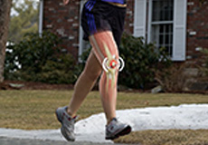
Knee Sports Injuries
Trauma is any injury caused during physical activity, motor vehicle accidents, electric shock, or other activities. Sports trauma or sports injuries refer to injuries caused while playing indoor or outdoor sports and exercising. Sports trauma can result from accidents, inadequate training, improper use of protective devices, or insufficient stretching or warm-up exercises.
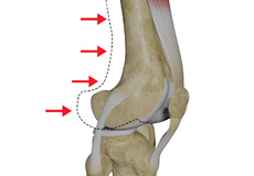
Tibial Plateau Fracture
A tibial plateau fracture is a crack or break on the top surface of the tibia or shinbone in the knee joint. The fracture most often occurs following a high-intensity trauma or injury from the impaction of the femoral condyles over the tibial plateau.
Patellar Tendinitis
Patellar tendinitis, also known as "jumper's knee", is an inflammation of the patellar tendon that connects your kneecap (patella) to your shinbone. This tendon helps in extension of the lower leg.
Women and ACL Injuries
The anterior cruciate ligament is one of the four major ligaments of the knee that connects the femur (thigh bone) to the tibia (shin bone) and helps stabilize the knee joint. Anterior cruciate ligament (ACL) injury is one of the common injuries of the knee.
Medial Meniscus Syndrome
Of the menisci within the knee, it is the medial that is more easily injured. Differences in the anatomical attachments of the medial meniscus compared to the lateral, mean that the medial meniscus becomes distorted during combined flexion and rotation movements in a manner not experienced on the lateral side.
Osgood Schlatter Disease
Osgood-Schlatter disease refers to a condition in older children and teenagers caused by excessive stress to the patellar tendon (located below the kneecap). Participants in sports such as soccer, gymnastics, basketball, and distance running are at higher risk for this disease.
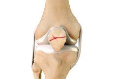
Tibial Eminence Fractures
The tibial eminence, also called the tibial spine, is a bony protuberance of the tibia (shin bone) that attaches to the anterior cruciate ligament (ACL) of the knee joint.
Anterior Knee Pain
Anterior knee pain is characterized by chronic pain over the front and center of the knee joint. It is common in athletes, active adolescents (especially girls) and overweight individuals. Anterior knee pain refers to various conditions, which include runner's knee or patellar tendinitis, and chondromalacia of the patella.
Runner's Knee
Patellofemoral pain syndrome also called runner’s knee refers to pain under and around your kneecap. Patellofemoral pain is associated with a number of medical conditions such as anterior knee pain syndrome, patellofemoral malalignment, and chondromalacia patella.
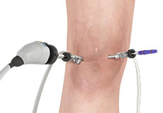
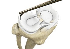
Knee Arthroscopy
Knee arthroscopy is a common surgical procedure performed using an arthroscope, a viewing instrument, to diagnose or treat a knee problem. It is a relatively safe procedure and you will usually be discharged from the hospital on the same day of surgery.
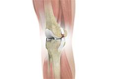
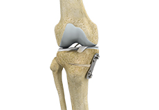
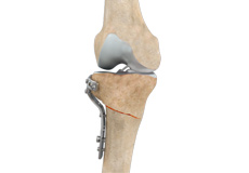
Knee Fracture Surgery
A knee fracture is a broken bone or a crack in or around the joint of the knee. This can involve the tibia (shin bone), the kneecap (patella), or femur (thighbone) where they connect with the knee.
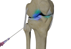
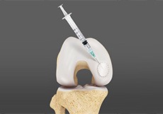
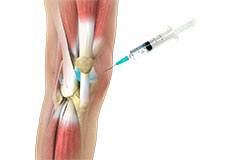
Intraarticluar Knee Injection
Knee pain and stiffness can be disabling and difficult to treat. It can limit an individual’s lifestyle and negatively impact body image and emotional well-being.
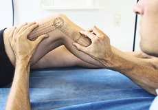
Physical Therapy for Knee
For knee problems, physical therapy involves strengthening and stretching certain joints and muscles. Physical therapy for the knee is designed to improve the muscle strength of the knee and reduce pain.
Posterolateral Corner (PLC) Reconstruction
A posterolateral corner is a complex arrangement of multiple ligaments, tendons, muscles and a joint capsule in your knee. It is located on the outside back corner of the knee.

Compartment Decompression
Compartment decompression, also called ‘decompressive fasciotomy’, is a surgical procedure to treat a painful knee condition known as “compartment syndrome”.
Revision Knee Ligament Reconstruction
Revision knee ligament reconstruction is a complex surgical procedure performed to address failures or to correct the undesirable consequences of primary reconstruction surgery on your knee.
Combined Hyaluronic Therapy for the Knee
Combined hyaluronic therapy is the process of injecting hyaluronic acid (HA) along with platelet-rich plasma (PRP) into your knee to treat osteoarthritis.
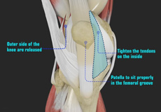
Patellofemoral Realignment
Patellofemoral realignment is a surgical procedure performed to treat symptomatic patellofemoral instability that does not respond to nonsurgical treatment measures.
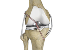
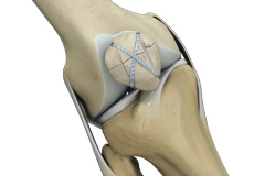
Knee Ligament Reconstruction
Knee ligament reconstruction is a surgical procedure to repair or replace damaged ligaments of the knee joint. The surgery can be performed using minimally invasive techniques.
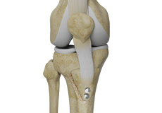
Tibial Tubercle Transfer
Tibial tubercle transfer is a surgical procedure that is performed along with other procedures to treat patellar instability, patellofemoral pain, and osteoarthritis. The tibial tubercle transfer technique involves realignment of the tibial tubercle (a bump in the front of the shinbone) such that the kneecap (patella) traverses in the center of the femoral groove.
Arthroscopic Debridement
Arthroscopic debridement or a clean-up is a surgical procedure performed using an arthroscope. In this procedure, the cartilage or the bone that is damaged is removed using surgical instruments and the edges of the articular cartilage that are rough will be smoothened.
LPFL Reconstruction
LPFL reconstruction or lateral patellofemoral ligament reconstruction is a surgical procedure employed to treat patients with severe patellofemoral instability. The procedure involves replacing a torn lateral patellofemoral ligament with a part of a tendon taken from your leg.
Failed Anterior Cruciate Ligament (ACL) Reconstruction
The knee joint is stabilized by four strong ligaments. The anterior cruciate ligament (ACL) passes diagonally in the middle of the knee, ensuring that the thigh and shin bone do not slide out of alignment during movement.
Failed Meniscus Repair
Meniscal repair may be performed either by open surgery under direct vision or minimally invasively using an arthroscope, which is a thin tube fitted with a camera that can be inserted into the knee through a very small incision to locate and repair the damaged meniscus.
Lateral Lengthening
Lateral lengthening, also known as lateral retinacular lengthening or release, is a surgical procedure to release a tightened lateral retinaculum on the outer aspect of the knee.
Meniscal Transplantation
Meniscal transplantation is a surgical procedure to replace the damaged meniscus of the knee with healthy cartilage.
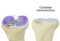
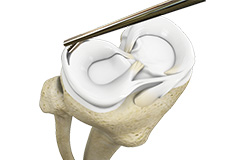
Meniscectomy
Meniscectomy is a surgical procedure indicated in individuals with torn meniscus where the conservative treatments are a failure to relieve the pain and other symptoms. Meniscectomy is recommended based on the ability of meniscus to heal, patient’s age, health status, and activity level.
Mosaicplasty
Weight-bearing joints, such as the knee, may develop defects in the articular cartilage (spongy tissue that lines and cushions joints during movement) due to stress, trauma or degenerative disease. This can lead to pain, swelling or locking at the joint.
Prior Meniscectomy
The menisci are two C-shaped cartilages that act as shock absorbers between the thigh and shin bones that articulate at the knee joint. They provide stability and lubrication to the joint as well as nutrition for the articular cartilage. Tears in the meniscus may occur as a result of acute injury or chronic degeneration with age.
Quadriceps Tendon Repair
Quadriceps tendon is a thick tissue located at the top of the kneecap. The quadriceps tendon works together with the quadriceps muscles to allow us to straighten our leg. The quadriceps muscles are the muscles located in front of the thigh.
Trochleoplasty
Before performing trochleoplasty, your doctor will ensure that the articular cartilage (spongy tissue that lines and cushions joints during movement) at the trochlea is normal and healthy. The surgery is avoided if there is any sign of arthritis.
Distal Femoral Osteotomy
An osteotomy is a surgical procedure that involves cutting of bone. The distal femur is part of the femur (thighbone) just above the knee joint. Distal femoral osteotomy is performed to correct knee alignment which can lead to excessive loading and degeneration of one side of the knee joint.
Hamstring Autograft
An autograft refers to using organ or tissue (bone, cartilage, tendon or skin) from the same person to transplant elsewhere on their body.

Pharmacological Interventions for Knee Injuries
Pharmacological interventions include medicinal preparations such as pain-relieving capsules or injections.
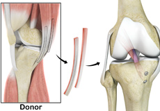
Hamstring Allograft
An allograft is an organ or tissue such as bone, cartilage, tendon or skin, taken from one person (donor) and surgically placed in another person to repair damaged tissue.
Viscosupplementation
Viscosupplementation refers to the injection of a hyaluronan preparation into the joint. Hyaluronan is a natural substance present in the joint fluid that assists in lubrication. It allows the smooth movement of the cartilage-covered articulating surfaces of the joint.
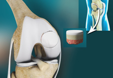
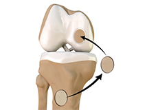
Knee Cartilage Restoration
Knee cartilage restoration is a surgical technique to repair damaged articular cartilage in the knee joint by stimulating new growth of cartilage or by transplanting cartilage into areas with defects in order to relieve pain and restore normal function to the knee.
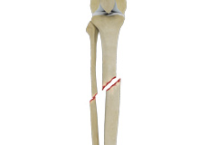
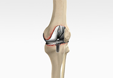
Knee Osteotomy
Knee osteotomy is a surgical procedure in which the upper shinbone (tibia) or lower thighbone (femur) is cut and realigned. It is usually performed in arthritic conditions affecting only one side of your knee. The aim is to take the pressure off the damaged area and shift it to the other side of your knee with healthy cartilage.
High Tibial Osteotomy
High tibial osteotomy is a surgical procedure performed to relieve pressure on the damaged site of an arthritic knee joint. It is usually performed in arthritic conditions affecting only one side of your knee and the aim is to take pressure off the damaged area and shift it to the other side of your knee with healthy cartilage.
Tibial Tubercle Osteotomy
Tibial tubercle osteotomy is a surgical procedure that is performed along with other procedures to treat patellar instability, patellofemoral pain, and osteoarthritis. The tibial tubercle transfer technique involves realignment of the tibial tubercle (a bump in the front of the shinbone) such that the kneecap (patella) traverses in the center of the femoral groove.
Multiligament Reconstruction of the Knee
Multiligament knee reconstruction is a surgical procedure to repair or replace two or more damaged ligaments of the knee joint. The surgery can be performed using minimally invasive techniques.
Patellar Tendon Repair
Patellar tendon repair is the surgery performed to reattach the torn tendon to the kneecap and to restore normal function in the affected leg.
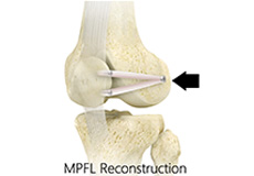
Medial Patellofemoral Ligament Reconstruction
The medial patellofemoral ligament can rupture or get damaged when there is patellar lateral dislocation. Dislocation can be caused by a direct blow to the knee, twisting injury to the lower leg, strong muscle contraction, or because of a congenital abnormality such as shallow or malformed joint surfaces.

Distal Realignment Procedures
Distal realignment procedures, also known as tibial tubercle transfer (TTT) procedures, are performed to reposition the kneecap after subluxation or dislocation by realigning the tendon under the kneecap to the underlying tibial tubercle.
PCL Reconstruction
PCL reconstruction surgery is a procedure to correct torn posterior cruciate ligament (PCL) in the knee using a tissue graft taken from another part of the body, or from a donor.
LCL Reconstruction
LCL reconstruction is a surgical procedure to repair torn or damaged lateral collateral ligament in the knee using a tissue graft taken from another part of the body, or from a donor.
ACL Reconstruction
ACL (anterior cruciate ligament) reconstruction is a commonly performed surgical procedure. With recent advances in arthroscopic surgery, it can now be performed with minimal incision and low complication rates.
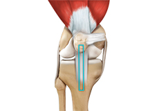
ACL Reconstruction with Patellar Tendon
Anterior cruciate ligament (ACL) reconstruction with patellar tendon is a surgical procedure that replaces the injured ACL with a patellar tendon. The goal of ACL reconstruction surgery is to tighten your knee and to restore its stability.
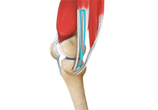
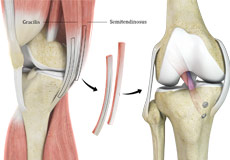
ACL Reconstruction Procedure with Hamstring Tendon
Anterior cruciate ligament (ACL) reconstruction with hamstring tendon is a surgical procedure to replace the torn ACL with part of the hamstring tendon taken from your leg. The hamstring is the muscle located on the back of your thigh. The goal of ACL reconstruction surgery is to tighten your knee and restore its stability.
MCL Reconstruction
MCL reconstruction is a minimally invasive surgical procedure in which a tendon graft is utilized to reconstruct the injured MCL.
Autologous Chondrocyte Implantation
Autologous chondrocyte implantation (ACI) is a procedure to treat the articular cartilage defects of the knee. This procedure is effective for treating small areas of cartilage damage that causes pain and swelling and restricts range of motion. Autologous chondrocyte implantation is not indicated if you have advanced arthritis of the knee.
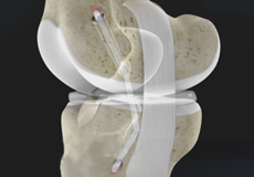
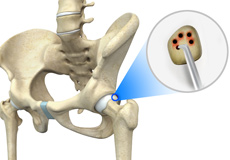
Subchondroplasty
Subchondroplasty is a minimally invasive procedure that is performed to specifically repair chronic BMLs by filling them with a bone substitute material. The bone substitute is then slowly resorbed and replaced with healthy bone, repairing the bone defect.
Partial Meniscectomy
Partial meniscectomy is a surgical procedure to remove the torn portion of the meniscus from the knee joint.
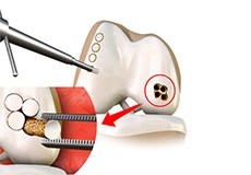
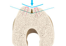
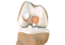
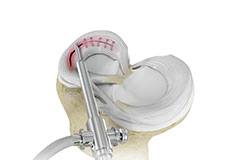
Cartilage Microfracture
Cartilage microfracture is a surgical procedure performed to replace the worn-out articular cartilage with new cartilage.
Meniscal Surgery
Meniscal surgery is a surgical procedure employed for the treatment of torn or damaged meniscal tissues in the knee. It is mostly performed as a minimally invasive keyhole procedure.
Transphyseal Surgery
ACL reconstruction via transphyseal surgery is recommended for active children who are involved in sports or recreational activities. Severe knee pain and inability to continue activities are the primary indications for ACL reconstruction.
Partial Transphyseal Surgery
Partial transphyseal surgery is an arthroscopically assisted operative procedure to reconstruct the anterior cruciate ligament (ACL) in the knee of a child or adolescent with growth plates or physis (areas near the ends of long bones where growth is still occurring).
Non-Surgical Knee Treatments
The knee is a complex joint which consists of bone, cartilage, ligaments, and tendons that make joint movements easy and at the same time it is more susceptible to various kinds of injuries. Knee problems may arise if any of these structures get injured by overuse or suddenly during sports activities.
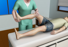
Physical Examination of the Knee
A complete physical examination of the knee is performed when you present to your doctor with a knee complaint. Both of your knees are examined and the results of the injured knee are compared to those of the healthy knee.

Pre-op and Post-op Knee Guidelines
Planning for your knee surgery prepares you for the operation and helps to ensure a smooth surgery and easier recovery. Here are certain pre-operative and post-operative guidelines which will help you prepare for knee surgery.

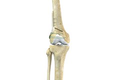
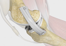
Am I a Candidate for Knee Surgery?
Arthritis of the knee can cause pain and stiffness, making regular activities such as walking and bending difficult. As arthritis progresses, conservative treatments tend to lose their efficacy and more definitive treatment should be considered. Knee replacement surgery involves replacing worn or damaged joints with implants to reduce pain and improve movement.
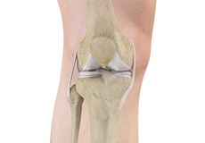
Knee Anatomy
The knee is a complex joint made up of different structures - bones, tendons, ligaments, and muscles. They all work together to maintain the knee’s normal function and provide stability to the knee during movement.
Having a well-functioning healthy knee is essential for our mobility and ability to participate in various activities. Understanding the anatomy of the knee enhances your ability to discuss and choose the right treatment procedure for knee problems with your doctor.
Bones of the Knee
The knee is a hinge joint made up of two bones, the thighbone (femur) and shinbone (tibia). There are two round knobs at the end of the femur called femoral condyles that articulate with the flat surface of the tibia called the tibial plateau. The tibial plateau on the inside of the leg is called the medial tibial plateau and on the outside of the leg, the lateral tibial plateau.
The two femoral condyles form a groove on the front (anterior) side of the knee called the patellofemoral groove. A small bone called the patella sits in this groove and forms the kneecap. It acts as a shield and protects the knee joint from direct trauma.
A fourth bone called the fibula is the other bone of the lower leg. This forms a small joint with the tibia. This joint has very little movement and is not considered a part of the main joint of the knee.
Articular Cartilage and Menisci of the Knee
Movement of the bones causes friction between the articulating surfaces. To reduce this friction, all articulating surfaces involved in the movement are covered with a white, shiny, slippery layer called articular cartilage. The articulating surface of the femoral condyles, tibial plateaus and the back of the patella are covered with this cartilage. The cartilage provides a smooth surface that facilitates easy movement.
To further reduce friction between the articulating surfaces of the bones, the knee joint is lined by a synovial membrane that produces a thick clear fluid called synovial fluid. This fluid lubricates and nourishes the cartilage and bones inside the joint capsule.
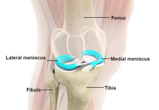
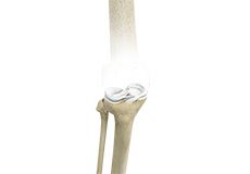
Within the knee joint, between the femur and tibia, are two C-shaped cartilaginous structures called menisci. Menisci function to provide stability to the knee by spreading the weight of the upper body across the whole surface of the tibial plateau. The menisci help in load-bearing i.e. it prevents the weight from concentrating onto a small area, which could damage the articular cartilage. The menisci also act as a cushion between the femur and tibia by absorbing the shock produced by activities such as walking, running and jumping.
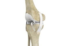
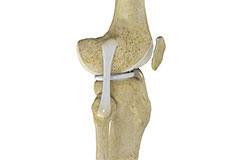
Ligaments of the Knee
Ligaments are tough bands of tissue that connect one bone to another bone. The ligaments of the knee stabilize the knee joint. There are two important groups of ligaments that hold the bones of the knee joint together, collateral and cruciate ligaments.
Collateral ligaments are present on either side of the knee. They prevent the knee from moving too far during side to side motion. The collateral ligament on the inside is called the medial collateral ligament (MCL) and the collateral ligament on the outside is called the lateral collateral ligament (LCL).
Cruciate ligaments, present inside the knee joint, control the back-and-forth motion of the knee. The cruciate ligament in the front of the knee is called anterior cruciate ligament (ACL) and the cruciate ligament in the back of the knee is called posterior cruciate ligament (PCL).
Muscles of the Knee
There are two major muscles in the knee - the quadriceps and the hamstrings, which enable movement of the knee joint. The quadriceps muscles are located in front of the thigh. When the quadriceps muscles contract, the knee straightens. The hamstrings are located at the back of the thigh. When the hamstring muscles contract, the knee bends.
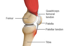
Tendons of the Knee
A tendon is a tissue that attaches a muscle to a bone. The quadriceps muscles of the knee meet just above the patella and attach to it through a tendon called the quadriceps tendon. The patella further attaches to the tibia through a tendon called the patella tendon. The quadriceps muscle, quadriceps tendon, and patellar tendon all work together to straighten the knee. Similarly, the hamstring muscles at the back of the leg are attached to the knee joint with the hamstring tendon.